A 50-year-old man visited the Comprehensive Headache Clinic at Bangkok Hospital Medical Center with the chief complaint of headaches occurring when he was laughing.
For the past thirty years, he had suffered from severe occipital pain provoked by laughing. He described the abrupt onset of sharp shooting pain at posterior part of upper cervical area, which radiated to bilateral occipital areas after vigorous laughing. The attacks lasted anywhere from a few seconds to a few minutes and were followed by a throbbing sensation at the same area which resolved spontaneously in less than an hour. The attacks only occured after laughing. Coughing, valsalva-like maneuver, straining when passing stool, change of position, or bending of neck could not precipitate headache or neck pain. He had no associated symptoms such as nausea, vomiting, photophobia, phonophobia, dizziness, fainting, blurring of vision, ptosis, lacrimation, conjunctival injection, or rhinorrhea during the attacks.
Bạn đang xem: VOLUME
Xem thêm : Picazón de la piel (prurito)
His past medical history was otherwise unremarkable. He denied history of head and neck injury, or chronic illness. Family history was unremarkable. No family members ever had headaches.
The neurological examination revealed normal cranial nerves function, normal motor power, no sensory loss, normal reflexes and normal gait. Neck examination showed mild restriction of range of motion both on flexion and extension, mild tenderness and spasm of upper posterior neck muscles, mild to moderate tenderness of occipital nerve area on both sides. Valsalva maneuver, coughing, and changing position (from lying to sit up and vice versa) was tried but could not provoke neck pain or headache.
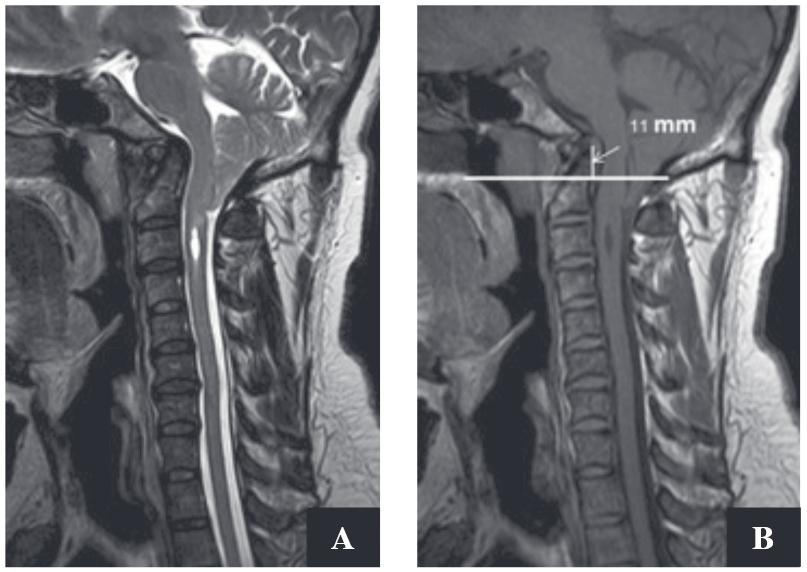
Secondary cause of headache and neck pain was suspected. MRI of cervical spine including the posterior fossa was performed. MRI demonstrated Arnold-Chiari type I malformation (ACM), syringomyelia at C-3 level, and mild intervertebral disc bulging (Figure 1).
Figure 1A-B: Sagittal T2W (A), and T1W (B) show downward displacement of the cerebellar tonsil at the posterior foramen magnum. There is high signal T2W of cavitary syrinx in cervical cord demonstrated in (A). The basilar invagination is also demonstrated as protruded dens with the tip at 11 mm. above theChamberlain line (B).
Neurosurgical consultation was done. Posterior fossa decompression was recommended but the patient refused the operation.
Nguồn: https://vuihoctienghan.edu.vn
Danh mục: Info
This post was last modified on Tháng mười một 20, 2024 12:25 sáng

