Where is it located?
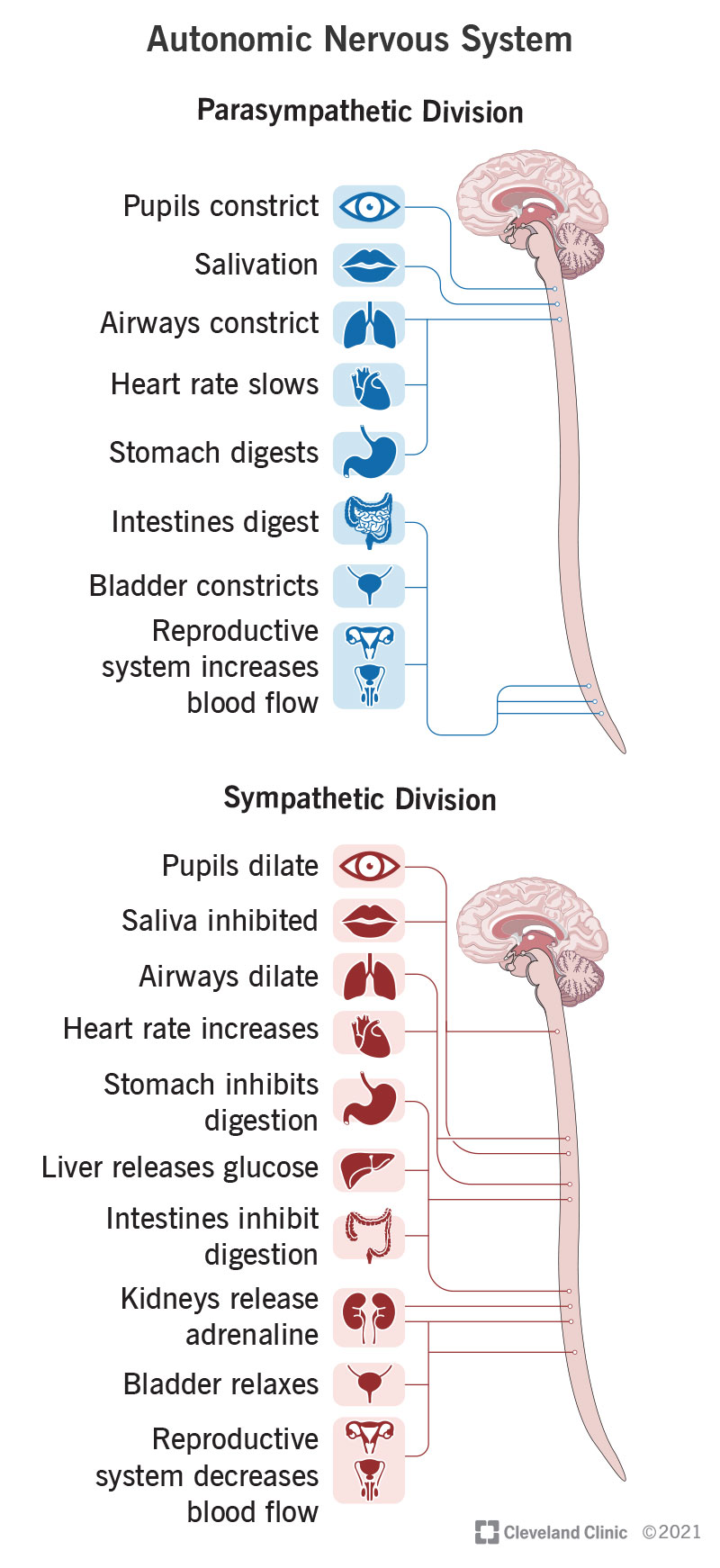
Your autonomic nervous system includes a network of nerves that extend throughout your head and body. Some of those nerves extend directly out from your brain, while others extend out from your spinal cord, which relays signals from your brain into those nerves.
There are 12 cranial nerves, which use Roman numerals to set them apart, and your autonomic nervous system has nerve fibers in four of them. These include the third, seventh, ninth and 10th cranial nerves. They manage pupil dilation, eye focusing, tears, nasal mucus, saliva and organs in your chest and belly.
Bạn đang xem: Autonomic Nervous System
Your autonomic nervous system also uses most of the 31 spinal nerves. These include spinal nerves in your thoracic (chest and upper back), lumbar (lower back) and sacral (tailbone).
Xem thêm : Antibodies
The spinal nerve connections are how your autonomic system controls the following:
- Heart.
- Lungs.
- Liver.
- Pancreas.
- Spleen.
- Stomach.
- Small and large intestine.
- Colon.
- Kidney.
- Bladder.
- Sexual organs.
The part of your brain that runs autonomic functions is your hypothalamus. This structure isn’t part of your autonomic nervous system, but is a key part of how it works.
What is it made of?
Your autonomic nervous system has a similar makeup to your overall nervous system. The main cell types are as follows, with more about them listed below:
- Neurons: These cells send and relay signals, and makeup parts of your brain, spinal cord and nerves. They also convert signals between the chemical and electrical forms.
- Glial cells: These cells don’t transmit or relay nervous system signals. Instead, they’re helpers or support cells for the neurons.
- Nuclei: These are nerve cell clusters grouped together because they have the same jobs or connections.
- Ganglia: These, pronounced “gang-lee-uh,” are groups of related nerve cells (one of these is a ganglion, pronounced “gang-lee-on”). They usually don’t all have the same jobs or connections, but they’re in roughly the same area or have connections to the same systems. Examples of this are the cochlear and vestibular ganglia, which are part of your senses of hearing and balance.
Neurons
Xem thêm : BLOG
Each neuron consists of the following:
- Cell body: This is the main part of the cell.
- Axon: This is a long, arm-like extension. At the end of an axon are several finger-like extensions where the electrical signal in the neuron becomes a chemical signal. These extensions, or synapses, connect with nearby nerve cells.
- Dendrites: These are small branch-like extensions (their name comes from a Latin word that means “tree-like”) on the cell body. Dendrites receive the chemical signals sent from synapses of other nearby neurons.
- Myelin: This is a thin layer composed of fatty compounds. Myelin is a protective covering that surrounds the axon of many neurons.
The dendrites on a single neuron may connect to thousands of other synapses. Some neurons are longer or shorter, depending on their location in your body and what they do.
Glial cells
Glial (pronounced “glee-uhl”) cells do several different jobs. They help develop and maintain neurons when you’re young and manage how neurons work throughout your life. They shield your nervous system from infections, control the chemical balance in your nervous system and coat neurons’ axons with myelin. There are 10 times more glial cells than neurons.
Nguồn: https://vuihoctienghan.edu.vn
Danh mục: Info


